Anatomy Of The Teeth Anatomical Chart
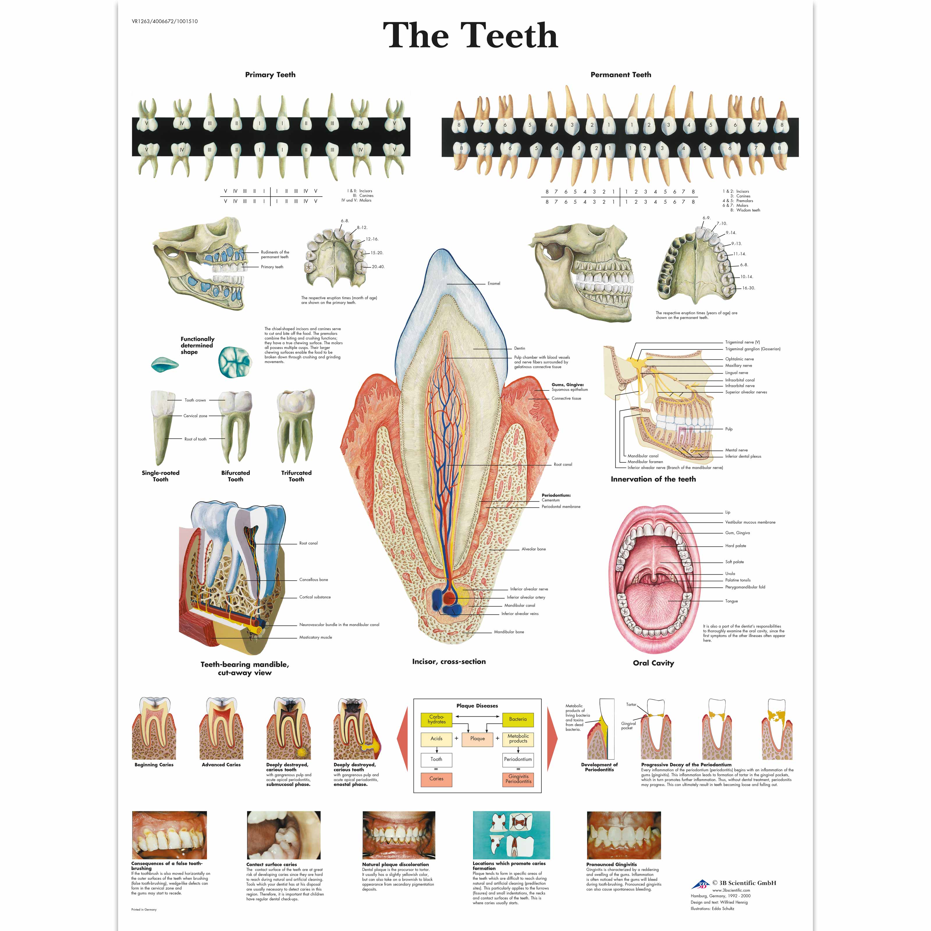
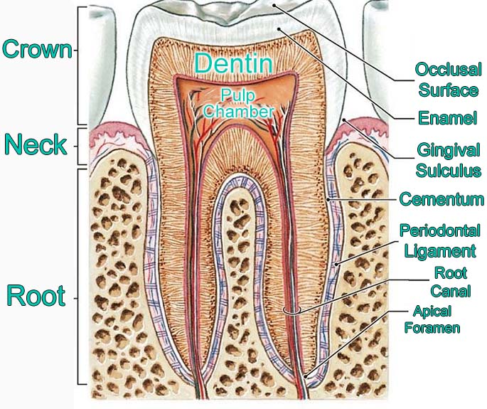
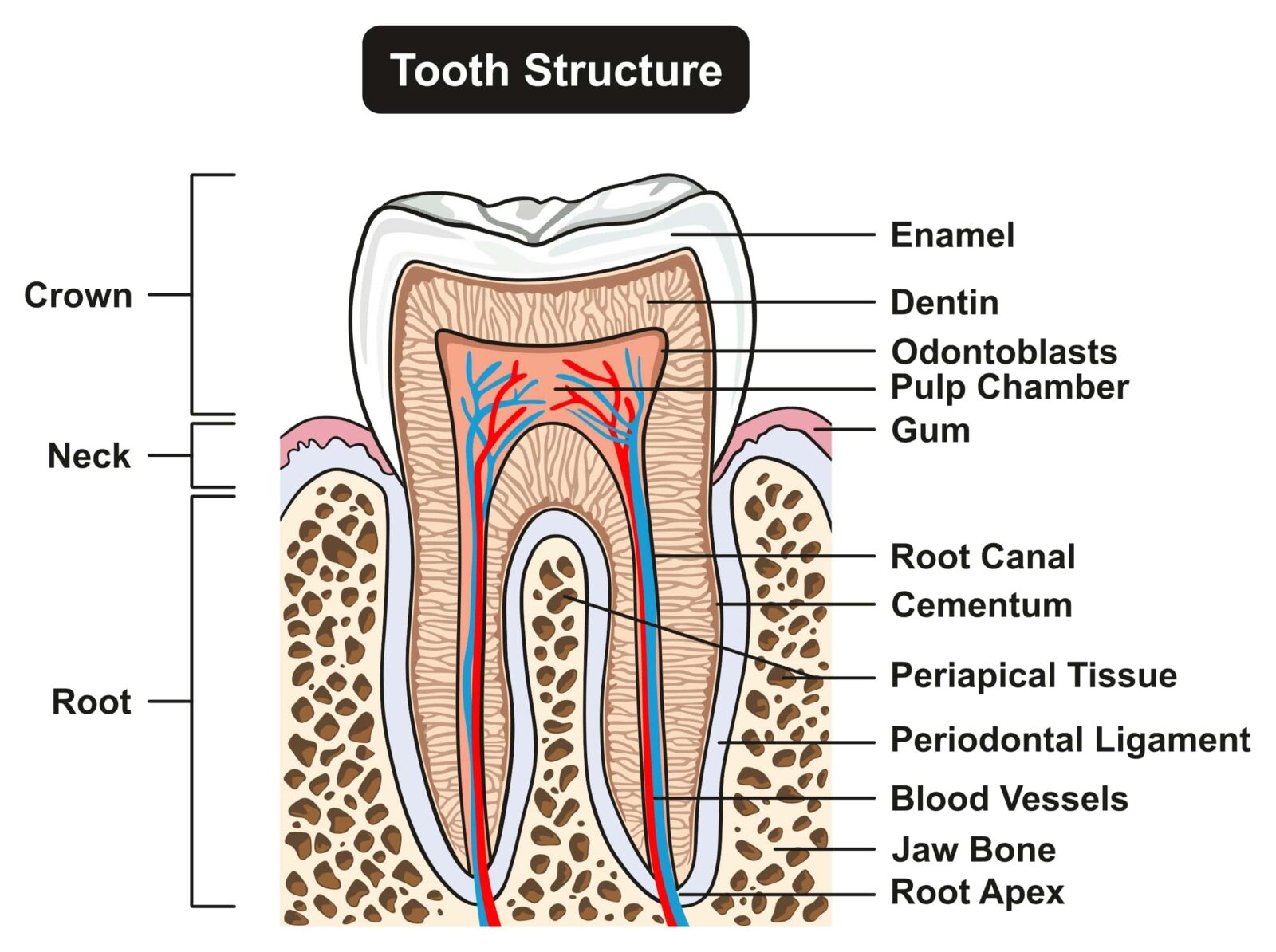
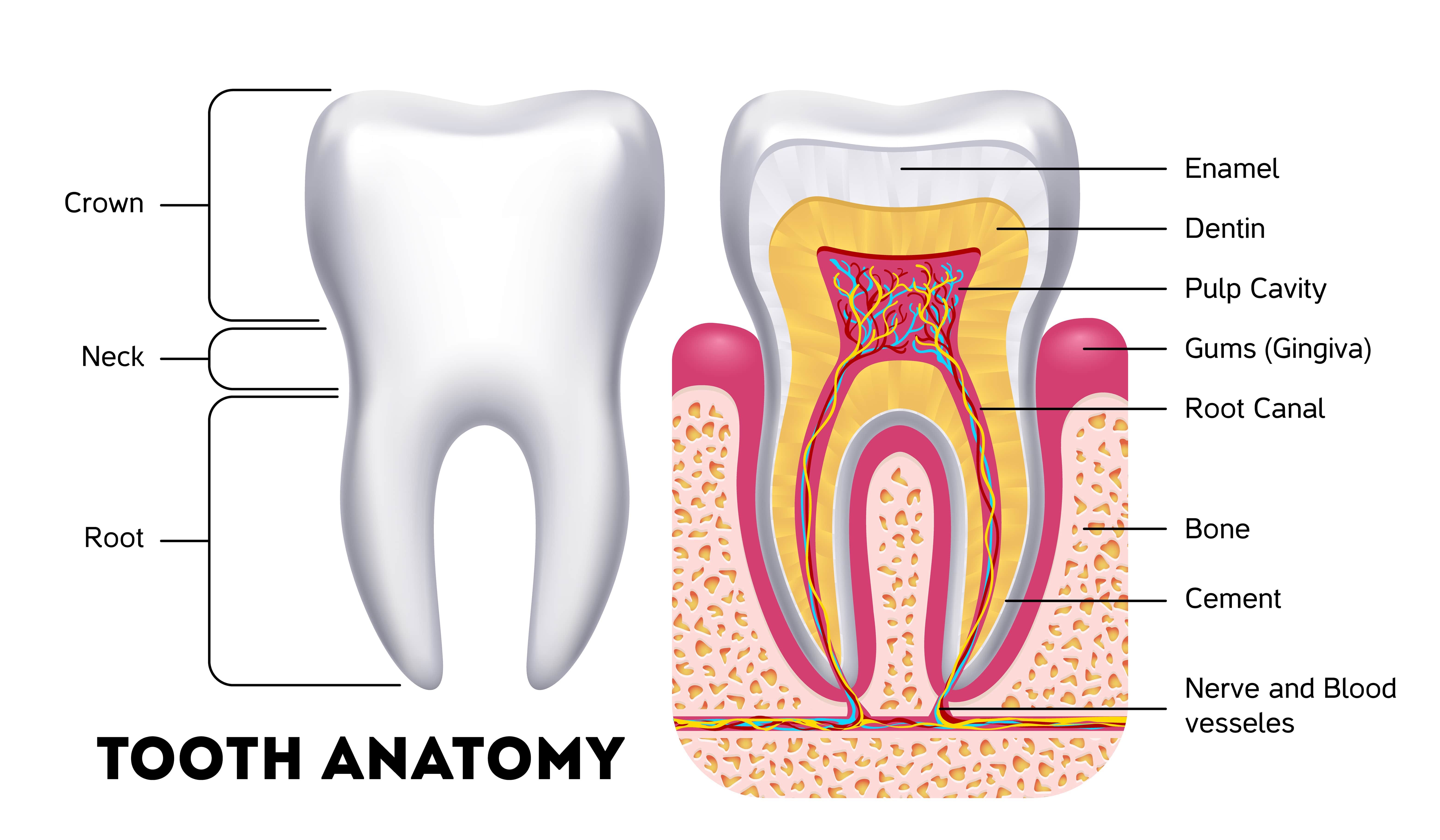
Anatomy Of The Teeth Anatomical Chart - The large central image shows a detailed cross section of a tooth and surrounding gum and bone with clearly labeled anatomic features. We’ll also go over some common conditions that can affect your teeth, and we’ll list common symptoms to watch for. This leaves up to eight adult teeth in each quadrant and separates the opposing pairs within the same alveolar bone as well as their counterparts in the opposing jaw. Though they look more like bones, teeth are actually ectodermal organs. Web in the tooth anatomy, we can find four types of teeth, each with a different job. Illustrates periodontal disease, three stages of dental caries, abscess formation, problems with the temporomandibular joint, glandular problems and impaction. This is the perfect diagram for any dental office, and colorful to grab a child's attention. The outside layer, called enamel, is. Teeth are positioned in alveolar sockets and connected to the bone by a suspensory periodontal ligament. Web the disorders of the teeth and jaw anatomical chart shows longitudinal section of a normal tooth. They cut and crush foods, making them easier to swallow. Web the teeth are categorized as incisors, canines, premolars, and molars and conventionally are numbered beginning with the maxillary right third molar (see figure identifying the teeth). Web learn about the types of teeth in a fast and efficient way using our interactive tooth identification quizzes and labeled diagrams. This leaves up to eight adult teeth in each quadrant and separates the opposing pairs within the same alveolar bone as well as their counterparts in the opposing jaw. The large central image shows a detailed cross section of a tooth and surrounding gum and bone with clearly labeled anatomic features. Tooth avulsion and enamel erosion). We’ll also go over some common conditions that can affect your teeth, and we’ll list common symptoms to watch for. Your teeth play a big role in digestion. Illustrates periodontal disease, three stages of dental caries, abscess formation, problems with the temporomandibular joint, glandular problems and impaction. Web anatomy of the teeth anatomical chart company staff,f. We’ll also go over some common conditions that can affect your teeth, and we’ll list common symptoms to watch for. Web anatomy now offers human 3d dental models and tooth charts as a means for educating patients on issues concerning the mouth, jaw, gums and teeth. Web the human teeth dental chart illustrates the types and working surfaces of the. Prefer to learn by doing? We’ll also go over some common conditions that can affect your teeth, and we’ll list common symptoms to watch for. Web the anatomy of a tooth divides into two main sections: Our mouths contain teeth of various shapes, sizes, and locations in the jaw. Web brightly colored, user friendly chart covering the anatomy of the. The crown and the root. Web each tooth consists of 3 anatomical parts: Web brightly colored, user friendly chart covering the anatomy of the teeth. The large central image shows a detailed cross section of a tooth and surrounding gum and bone with clearly labeled anatomic features. Web we’ll go over the anatomy of a tooth and the function of. Web anatomy of the teeth anatomical chart company staff,f. Most people have 32 teeth, but that can vary. Web learn about the types of teeth in a fast and efficient way using our interactive tooth identification quizzes and labeled diagrams. The numbering system shown is the one most commonly used in the united states. Web the four main types of. Web most adults have 32 permanent teeth, including eight incisors, four canines, eight premolars and 12 molars. How teeth are shaped and aligned affect your smile, speech, and facial shape. Teeth are positioned in alveolar sockets and connected to the bone by a suspensory periodontal ligament. The outside layer, called enamel, is. Our mouths contain teeth of various shapes, sizes,. Web each tooth consists of 3 anatomical parts: Teeth are positioned in alveolar sockets and connected to the bone by a suspensory periodontal ligament. Most people have 32 teeth, but that can vary. Also includes labeled illustrations of the following: The crown, neck, and root. Web we’ll go over the anatomy of a tooth and the function of each part. Web brightly colored, user friendly chart covering the anatomy of the teeth. The crown and the root. Web brightly colored, user friendly chart covering the anatomy of the teeth. Also includes labeled illustrations of the following: They cut and crush foods, making them easier to swallow. Each type of tooth is designed to perform different functions, like biting, tearing, and chewing. Web in the tooth anatomy, we can find four types of teeth, each with a different job. Web the teeth are categorized as incisors, canines, premolars, and molars and conventionally are numbered beginning with the. The numbering system shown is the one most commonly used in the united states. It is the visible portion of the tooth that protrudes from the gum. Also includes labeled illustrations of the following: Web depicted is the relationships of the 32 teeth of the human mouth and the internal organs they affect, including the vascular, digestive, and respiratory systems.. There are dental charts showing disorders of the jaw and other diseases of the dental structure. Look no further than our dental anatomy quizzes and tooth diagrams. Web the teeth are categorized as incisors, canines, premolars, and molars and conventionally are numbered beginning with the maxillary right third molar (see figure identifying the teeth). Fully labeled illustrations of the teeth. Also includes labeled illustrations of the following: They cut and crush foods, making them easier to swallow. The large central image shows a detailed cross section of a tooth and surrounding gum and bone with clearly labeled anatomic features. Web brightly colored, user friendly chart covering the anatomy of the teeth. The crown, neck, and root. Web anatomy of the teeth anatomical chart company staff,f. Web brightly colored, user friendly chart covering the anatomy of the teeth. Your teeth play a big role in digestion. It is the visible portion of the tooth that protrudes from the gum. Web we’ll go over the anatomy of a tooth and the function of each part. Web the anatomy of a tooth divides into two main sections: The large central image shows a detailed cross section of a tooth and surrounding gum and bone with clearly labeled anatomic features. Web in the tooth anatomy, we can find four types of teeth, each with a different job. Web most adults have 32 permanent teeth, including eight incisors, four canines, eight premolars and 12 molars. Web each tooth consists of 3 anatomical parts: The outside layer, called enamel, is.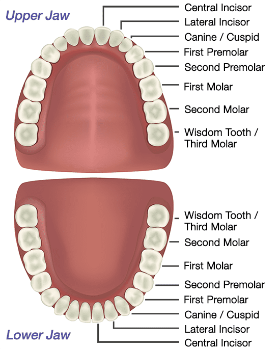
Tooth Anatomy Gosford, Experienced Dentists VC Dental
Anatomical Charts and Posters Anatomy Charts Dental Charts The
Parts Of A Tooth Anatomy

Dental Anatomy Chart Teeth Jaw Poster Tooth Anatomical

Tooth Anatomy Poster Behance

Teeth Anatomical Chart

Anatomy of the Teeth Anatomical Chart — Australia
What's Inside Your Teeth? Acorn Dentistry For Kids
Anatomy Of The Teeth Anatomical Chart Poster Prints Images and Photos

Dental Anatomy Study Guide (8.5" X 11") 3Panel, Laminated
Tooth Avulsion And Enamel Erosion).
Our Mouths Contain Teeth Of Various Shapes, Sizes, And Locations In The Jaw.
Fully Labeled Illustrations Of The Teeth With Dental Terminology (Orientation, Surfaces, Cusps, Roots Numbering Systems) And Detailed Images Of Each Permanent Tooth.
The Large Central Image Shows A Detailed Cross Section Of A Tooth And Surrounding Gum And Bone With Clearly Labeled Anatomic Features.
Related Post: